Illuminating the Inner Retina of Vertebrates: Multiple Opsins and Non-Visual Photoreceptors
REVIEW
Abstract
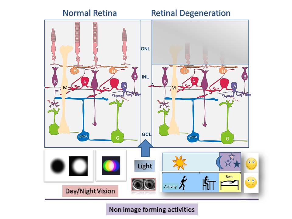
Throughout evolution, the need to detect light has generated highly specialized photoreceptor cells that in vertebrates are mainly located in the retina. The most studied photodetectors within these cells are the visual photoreceptors “cones and rods” responsible for day and night vision, respectively. These cells contain photosensitive molecules consisting of a protein part called “opsin” that binds a chromophore derived from vitamin A, retinaldehyde, capable of photoisomerizing from 11-cis retinal to all-trans retinal form, and triggering the light responses that lead to vision. However, other cells of the inner retina of vertebrates [retinal ganglion cells (RGCs), horizontal cells (HCs), and Muller’s glial cells] are currently known to express non-visual photopigments such as melanopsin (Opn4), encephalopsin (Opn3) and neuropsin (Opn5), which would be involved in diverse functions not associated with imaging. Melanopsin is the most widely studied of them, it is expressed in intrinsically photosensitive RGCs (ipRGCs) and HCs of the chicken retina and participates in setting the biological clock, the pupillary light reflex, and presumably in other reflex and subconscious functions, in addition to the lateral interaction between visual photoreceptors and HCs. It is noteworthy that these non-visual photopigments (Opn3, Opn4 and Opn5) respond to blue and/or near violet region light. This particular photosensitivity may provide individuals with a broader spectrum of response to light stimulation within the visible beyond the scope of the visual photoreceptors, regulating an important number of functions not yet completely identified. We can conclude that “a constellation of cells and photoreceptor molecules are present in the inner retina of vertebrates, and from very early stages of development, even before any sign of vision may occur.”


